Hip Anatomy
The hip joint is the biggest joint in the body that supports body weight. It is a type of ball-and-socket joint and is protected by muscles, ligaments, and tendons. The hip joint is created at the point where the thigh bone, known as the femur, joins the pelvis.
If the hip joint is injured or becomes ill, it can limit how much the joint can move and make it harder for the body to support weight.
The hip joint is made up of the following:
- Bones and joints
- Ligaments of the joint capsule
- Muscles and tendons
- Nerves and blood vessels that supply the bones and muscles of the hip
Bones and Joints
The hip joint is the point where the leg connects to the trunk of the body. It is made up of two main bones: the femur (thigh bone) and the pelvis, which consists of three bones called the ilium, ischium, and pubis. The femoral head forms the “ball” of the hip joint, while the “socket” is created by the acetabulum. The acetabulum is a deep, circular cavity located on the outer edge of the pelvis, formed by the fusion of the ilium, ischium, and pubis. The lower part of the ilium connects with the pubis, and the ischium lies behind it. The hip joint is stabilized by the acetabulum, surrounding muscles, ligaments, and the joint capsule.
Within the acetabulum, the femoral head rotates and glides, allowing a wide range of movement. A fibrocartilaginous rim called the labrum is attached to the acetabulum, which deepens the socket and enhances joint stability.
The femur, or thigh bone, is among the longest bones in the human body. Its upper section includes the femoral head, femoral neck, and two bony projections called the greater and lesser trochanters, which serve as attachment points for muscles. The femoral head fits into the acetabulum of the pelvis to form the hip joint.
Covering the surfaces of weight-bearing bones is a thin, durable layer of articular cartilage, lubricated by synovial fluid. This cartilage ensures smooth movement of the bones and minimizes friction within the joint.
Ligaments
Ligaments are strong, fibrous bands that link one bone to another. Around the hip joint, several ligaments form a firm outer layer over the joint capsule, providing strength and stability. The main ligaments connected to the hip joint include:
- Iliofemoral ligament – This is a Y-shaped ligament that connects the pelvis to the front portion of the hip joint, specifically the femoral head. It helps to limit how far the hip can extend.
- Pubofemoral ligament – This triangular-shaped ligament extends from the upper portion of the pubis and attaches to the iliofemoral ligament. It provides a connection between the pubis and the femoral head.
- Ischiofemoral ligament – This is a group of strong fibers that originate from the ischium, located behind the acetabulum, and combine with the fibers of the joint capsule.
- Ligamentum teres – This is a small ligament that runs from the end of the femoral head to the acetabulum. Even though it does not play a role in moving the hip, it contains a small blood vessel that provides blood to part of the femoral head.
- Acetabular labrum – The labrum is a ring of fibrous cartilage that surrounds the acetabular socket. It makes the socket deeper, which helps to improve the stability and strength of the hip joint.
Muscles and Tendons
A long tendon known as the iliotibial band runs along the femur from the hip to the knee and acts as a point where several hip muscles attach, including the following:
- Gluteals – The muscles that make up the buttocks consist of three specific muscles: the gluteus minimus, gluteus maximus, and gluteus medius. These muscles connect to the back of the pelvis and attach to the greater trochanter of the femur.
- Adductors – These muscles are found in the thigh and assist in adduction, which is the movement of pulling the leg back toward the center of the body.
- Iliopsoas – This muscle lies at the front of the hip joint and plays a key role in flexing the leg at the hip. It is a deep muscle that begins in the lower back and pelvis and extends to attach on the inner surface of the upper femur.
- Rectus femoris – This is the biggest group of muscles found in front of the thigh area. They also function as hip flexors.
- Hamstring muscles – These muscles originate from the lower region of the pelvis and extend down the back of the thigh. Since they pass behind the hip joint, their main function is to pull the hip backward, a motion known as hip extension.
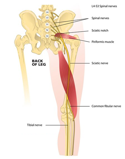
Nerves and Arteries
The nerves located in the hip area carry messages from the brain to the muscles, which helps regulate the movement of the hip. They also send sensory information, such as touch, pain, and temperature, back to the brain.
A smaller nerve known as the obturator nerve also supplies the hip. The main nerves found in the hip area are the femoral nerve, which is located in front of the thigh bone, and the sciatic nerve, which is found at the back.
Along with the nerves, there are blood vessels that provide blood to the lower limbs. The femoral artery, one of the largest arteries in the body, starts deep within the pelvis and can be felt in front of the upper part of the thigh.
Hip movements
The different parts of the hip work together to allow for various types of movement. The movements of the hip include bending forward (flexion), moving backward (extension), moving away from the body (abduction), moving toward the body (adduction), moving in a circular motion (circumduction), and rotating the hip.