Foot and Ankle Anatomy
Introduction
The foot and ankle work together in the human body to help with balance, stability, movement, and pushing off during walking or running.
This complex anatomy consists of:
- 26 bones
- 33 joints
- Muscles
- Tendons
- Ligaments
- Blood vessels, nerves, and soft tissue
To better understand conditions that affect the foot and ankle, it is important to know how the normal structure of the foot and ankle is built.
Ankle
The ankle is made up of three bones that are linked together by muscles, tendons, and ligaments. These structures help connect the foot to the leg.
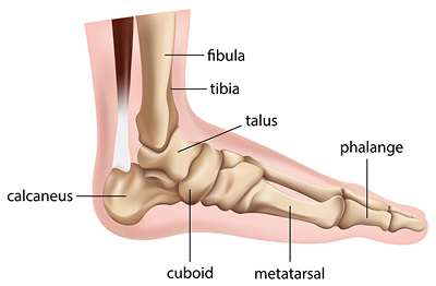
The lower leg consists of two primary bones: the tibia, or shin bone, and the fibula. They join with the talus, or ankle bone, at the tibiotalar joint. This joint, commonly referred to as the ankle joint, makes the up-and-down movement of the foot possible.
- Tibia (shin bone)
- Fibula
- Talus
- Lateral Malleolus
- Ligaments
- Medial Malleolus
The bony protrusions that we can see and feel on the ankle are:
- Lateral Malleolus: The outer ankle bone is made up of the lower part of the fibula.
- Medial Malleolus: The inner ankle bone is formed by the lower part of the tibia.
Hindfoot
The foot is divided into three main parts known as the hindfoot, midfoot, and forefoot. The hindfoot includes the talus bone, which is also referred to as the ankle bone, and the calcaneus bone, which is commonly known as the heel bone. The calcaneus is the largest bone in the foot, and the talus is the highest bone in that area. These two bones are joined by the subtalar joint, which lets the foot turn at the ankle.
The hindfoot connects the midfoot to the ankle at the transverse tarsal joint.
- Talus
- Calcaneus
Midfoot
The midfoot is made up of five tarsal bones, including the navicular bone, the cuboid bone, and the three cuneiform bones. It links the front part of the foot to the back part, and is supported by muscles and ligaments. The most important ligament in this area is the plantar fascia. The midfoot is essential for creating the foot's arches and helps absorb shocks during activities like walking or running.
The midfoot is joined to the forefoot through five tarsometatarsal joints.
- Navicular
- Cuboid
- Cuneiform Bones
Forefoot
The forefoot consists of the metatarsals, which are the long bones in the foot, and the phalanges, which make up the toes. These parts are connected at the ball of the foot through the metatarsophalangeal joints. Each of the smaller toes has three phalanges and two joints, whereas the big toe has two phalanges, two joints, and also includes two small sesamoid bones. These sesamoid bones are located within a tendon close to the joint and help support the movement of the big toe, allowing it to bend up and down.
The first metatarsal bone, which connects to the big toe, is the shortest and thickest among the metatarsal bones. It serves as an attachment point for multiple tendons. This bone plays a key role in pushing the body forward during movement and supporting the body's weight.
- Phalanges
- Metatarsal
Soft Tissue Anatomy
Different types of soft tissues play a role in supporting and maintaining the stability of the bones in the feet and ankles.
- Cartilage: Cartilage is shiny and smooth, helping bones glide easily and allowing joints to move without friction.
- endons: Tendons are types of soft tissue that link muscles to bones, helping to support the body. The Achilles tendon, also known as the heel cord, is the biggest and strongest tendon in the body. It is found on the back of the lower leg and runs around the calcaneus, which is the heel bone. If this tendon becomes inflamed, it can lead to a painful condition called Achilles tendonitis, which can make walking very difficult because of the discomfort it causes.
- Ligaments: Ligaments are strong, rope-like tissues that connect bones and help keep joints stable by supporting tendons. The plantar fascia is the longest ligament in the foot. It begins at the heel bone, known as the calcaneus, and runs along the bottom of the foot toward the front. It helps support the arches of the foot and also helps absorb shock when you move. Plantar fasciitis is a common issue that causes pain in the heel of adults. It occurs when repeated stress causes small tears in the plantar fascia. Another common injury is an ankle sprain, which happens when ligaments are stretched too much or torn. This usually affects the talo-fibular and calcaneo-fibular ligaments.
- Muscles: Muscles are fibrous tissue that can contract, enabling the body to move. The foot contains 20 muscles, which are categorized into two main groups: intrinsic and extrinsic. The intrinsic muscles lie within the foot and control toe movements. The extrinsic muscles, on the other hand, are situated in the lower leg and are positioned outside the foot. The gastrocnemius, also known as the calf muscle, is the largest of these and helps in moving the foot. Muscle strains often happen when a muscle is overused, especially if it is stretched without being properly warmed up.
- Bursae: Bursae are small sacs filled with fluid that help reduce friction between tendons, bones, and skin. These sacs contain specific cells known as synovial cells, which produce a lubricating fluid. If this fluid becomes infected, it can lead to a common and painful condition called Bursitis.
Biomechanics of Foot & Ankle
Biomechanics is a term to describe movement of the body. The ankle joint by itself permits two movements:
- Plantar flexion:Pointing the foot downward. This action is usually done along with turning the sole of the foot inward.
- Dorsiflexion: Lifting the foot upward. This movement is typically paired with turning the sole of the foot outward.
The foot (excluding the toes) also permits two movements:
- Inversion: Turning the sole of the foot inward.
- Eversion: Turning the sole of the foot outward
The toes allow four different movements:
- Plantar flexion: Curling the toes down toward the bottom of the foot.
- Dorsiflexion: Bending the toes towards the top of the foot
- Abduction: Spreading the toes apart. This movement normally accompanies plantar dorsiflexion.
- Adduction: Bringing the toes together. This movement normally accompanies plantar flexion.